Gynec Imaging in Ahmedabad

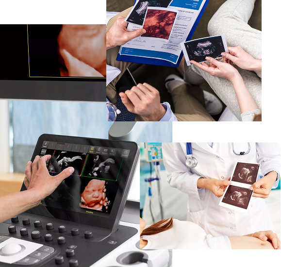
Accurate Ultrasound & Imaging for Women’s Health

Excel Hospital in Ahmedabad offers advanced gynecological imaging services designed to assess and diagnose a wide range of women’s health concerns. Our imaging facilities include high-resolution ultrasound (2D, 3D, and Doppler) and specialized pelvic scans to examine the uterus, ovaries, fallopian tubes, and surrounding pelvic structures. These tests help in the early detection of conditions such as fibroids, ovarian cysts, endometriosis, and pregnancy-related complications.

With modern imaging equipment, expert radiologists, and strict medical protocols, we ensure every scan is accurate, safe, and comfortable for our patients. Whether it’s a routine pelvic ultrasound, obstetric scan, or Doppler blood flow study, our goal is to provide clear results that support precise diagnosis and effective treatment planning.

Our Services

Imaging for accurate gynecological diagnosis.

At Excel Hospital, we provide precise diagnostic imaging for women’s health needs. Using advanced ultrasound and Doppler technology, our team ensures clear visualization and accurate reporting for gynecological and obstetric conditions.
Pelvic ultrasound
Transvaginal scan
Obstetric ultrasound
Follicular study
3D/4D ultrasound
Pelvic Doppler
Endometrial scan
Ovarian evaluation
Fibroid assessment
USG-guided biopsy
Dr. Aarti Vazirani’s

Expertise in Gynecological Imaging

Dr. Aarti Vazirani brings over 15 years of clinical experience in women’s health diagnostics. At Excel Hospital’s Gynec Imaging unit, she performs and interprets advanced pelvic, obstetric, and Doppler scans to support timely and accurate gynecological care. Her training in high-resolution and transvaginal imaging ensures clear visualization of complex pelvic structures.

Her patient-centered approach emphasizes comfort, clear communication, and precise results. With expertise in pregnancy monitoring, infertility workups, and early detection of gynecological conditions, Dr. Vazirani’s imaging interpretations play a vital role in guiding effective treatment plans in collaboration with our multidisciplinary team.

Our Testimonials

Patient feedback on gynec imaging care.

FAQs

Gynecological ultrasound uses high-frequency sound waves to create real-time images of pelvic organs such as the uterus, ovaries, and fallopian tubes. It helps detect structural changes, monitor pregnancy, and evaluate conditions like fibroids, cysts, or endometrial abnormalities without using radiation.
A pelvic ultrasound is performed over the lower abdomen and is often used for an overview of pelvic organs. A transvaginal scan uses a thin probe inserted into the vagina, offering a closer and more detailed view of the uterus, ovaries, and surrounding tissues, especially useful for early pregnancy or subtle abnormalities.
Doppler ultrasound evaluates blood flow in pelvic vessels, ovarian masses, or the placenta during pregnancy. It is particularly important in high-risk pregnancies, suspected ovarian torsion, or to assess the vascularity of pelvic tumors.
While ultrasound is excellent for visualizing structural abnormalities, some conditions like very early-stage endometriosis or microscopic lesions may require additional tests such as MRI, CT, or hysteroscopy for confirmation. Your doctor will decide if further imaging is needed.
Since ultrasound does not use radiation, it can be repeated as often as needed for monitoring ongoing conditions or pregnancy. However, unnecessary scans are avoided, and imaging is recommended only when clinically appropriate.